Le Club industrie 4.0 :
Créé en 2018 par Robotics Valley et l’UIMM Côte-d’Or, le Club Industrie 4.0 offre l’opportunité de rencontres et d’échanges entre industriels issus de secteurs d’activité et de typologies d’entreprises différents. Ce club est ouvert à toutes les entreprises industrielles adhérentes à Robotics Valley et de ses partenaires.
Présent à Dijon depuis sa création, le club industrie s’est étendu à Chalon-sur-Saône / Le Creusot. Et bientôt dans d’autres lieux de la région Bourgogne Franche-Comté.
Les thématiques abordées sont variées et en rapport direct avec les problématiques rencontrées dans le quotidien des industriels présents (l’automatisation robotisée des processus (RPA), No-Code et Low-Code, l’intelligence artificielle et les usages pour l’industrie).
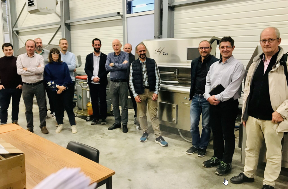
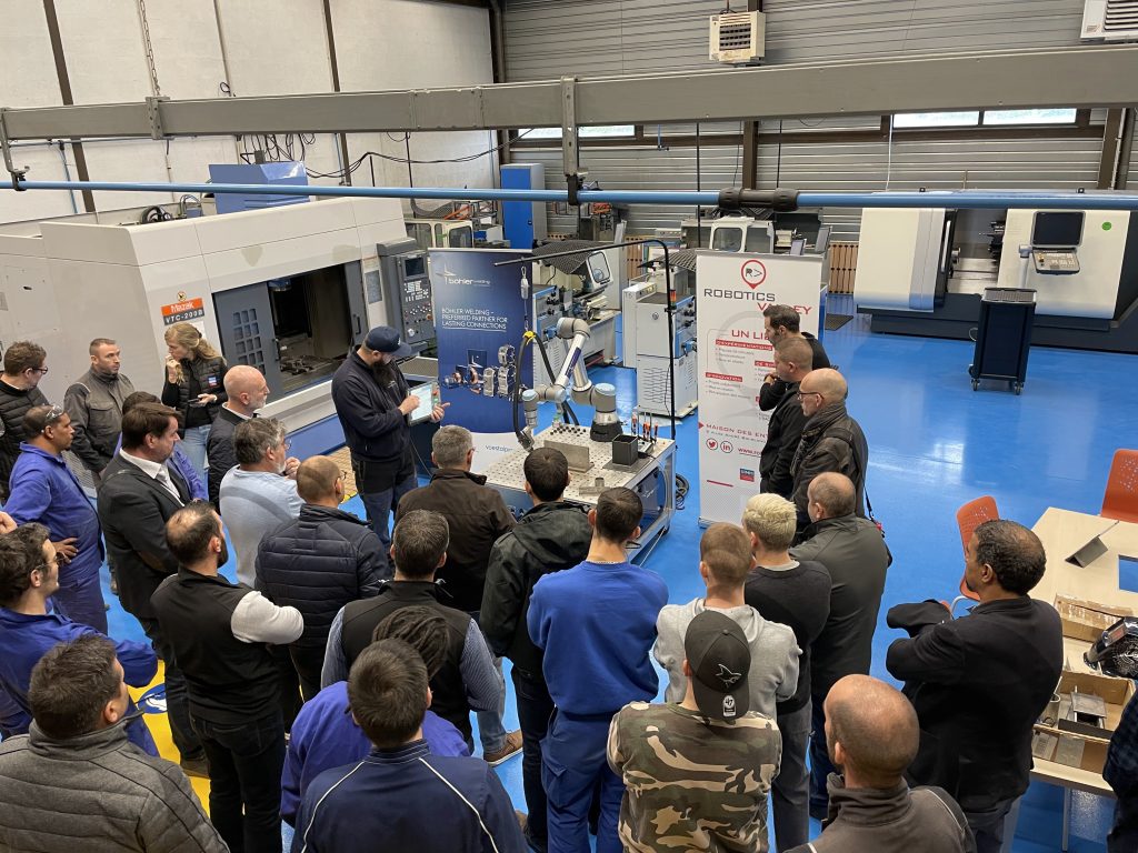
Les journées techniques :
Ces journées ont pour but d’acculturer les industriels aux technologies de l’industrie 4.0. Elles s’articulent autour de présentations et de démonstrations, durant lesquelles les participants peuvent manipuler les outils présentés.
Les journées techniques peuvent porter sur différentes thématiques telles que l’IoT, le soudage ou la robotique…
Les groupes de travail :
Les groupes de travail sont basés sur des thématiques communes et réalisés en partenariat avec des acteurs majeurs de l’industrie et des institutionnels.
- Réflexion sur l’écosystème 4.0 sur la région Bourgogne-Franche-Comté pour rendre visible le territoire et ses compétences afin d’attirer des talents et créer des synergies entre les entreprises technologiques et industrielles ;
- Rencontres entre intégrateurs pour permettre des échanges et retours d’expériences sur des problématiques communes.

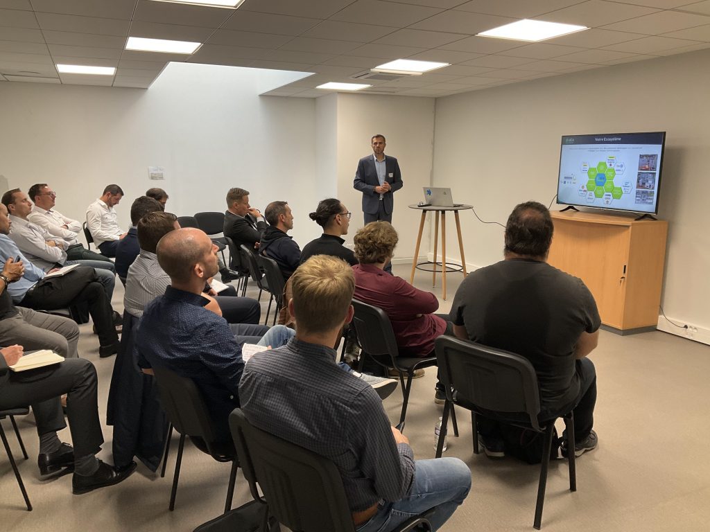
Les Webinaires :
Chaque dernier mardi du mois, nous organisons un webinaire en association avec l’une de nos entreprises adhérentes. L’objectif de ces webinaires est de mettre en avant cette entreprise ainsi que le produit ou la solution qu’elle souhaite promouvoir.
Chaque mardi du mois, une entreprise, une solution innovante à découvrir via les éditions des webinaires mensuels de Robotics Valley.
Les journées, rencontre labos – industriels :
L'objectif est de créer des ponts entre les industriels et les laboratoires et d'imaginer des collaborations.
Cette rencontre offre l'opportunité d'échanger en face à face et en toute discrétion avec les chercheurs, afin de discuter de vos problématiques de R&D et de projets d'innovation.